Figure 1.5 from Sharpe, L. T., Stockman, A., Jägle, H., & Nathans, J. (1999). Opsin genes, cone photopigments, color vision and colorblindness. In K. Gegenfurtner & L. T. Sharpe (Eds.), Color vision: From Genes to Perception (pp. 3-51) Cambridge: Cambridge University Press.
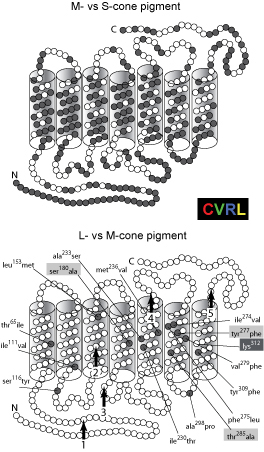
Cone opsin amino acid differences. Pairwise comparisons of human visual pigment molecules showing amino-acid identities (open circles) and differences (filled circles) (after Nathans, Thomas, & Hogness, 1986). In each representation, the seven α-helices are arranged in a line. When intramembrane regions are optimally aligned, the amino-proximal tails (extracellular face) of the M- (or L-) cone pigments are 16 amino acids longer than for the S-cone pigment. The alignment can be improved by inserting into the M- (or L-) cone pigment sequences gaps of two amino acids and of one amino acid, respectively, at positions 4 residues and 29 residues from the carboxyl terminus. (Top) Identity between the M- and S-cone pigments. (Bottom) Identity between the L- and M-cone pigments. The location of lysine312, the site of covalent attachment of 11-cis retinal, and the 15 amino acid substitutions are indicated. The start of each of the 5 intron positions are indicated by numbered vertical arrows. The subsitutions at codons 180, 277, and 285 (highlighted) are believed to contribute the majority of the spectral difference between the M- and L-cone pigments.
Nathans, J., Thomas, D., & Hogness, S. G. (1986). Molecular genetics of human color vision: The genes encoding blue, green and red pigments. Science, 232, 193-202.
Sharpe, L. T., Stockman, A., Jägle, H., & Nathans, J. (1999). Opsin genes, cone photopigments, color vision and colorblindness. In K. Gegenfurtner & L. T. Sharpe (Eds.), Color vision: From Genes to Perception (pp. 3-51) Cambridge: Cambridge University Press.