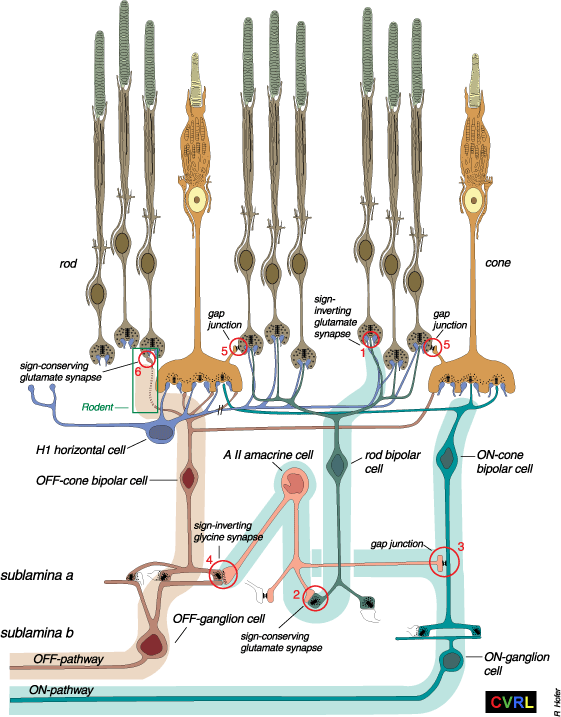

Figure 1 from Sharpe, L. T., & Stockman, A. (1999). Two rod pathways: the importance of seeing nothing. Trends in Neurosciences, 22, 497-504.
Rod and cone pathways in the mammalian retina. The retina is a complex neural tissue interweaving multiple circuits for transmitting photon signals from the light-sensitive rod and cone photoreceptors to the ON- and OFF-ganglion cells, the axons of which form the optic nerve. Integral to the circuits are bipolar, amacrine and horizontal cells, which maintain or enhance the linkage. The highly schematic retinal diagram depicted here concentrates on the pathways available to the rods, all of which either infiltrate or superimpose upon the cone circuitry (see Box 1). The numbered circles highlight the six so-far identified or inferred regions of rod-signal transmission: (1) the rod-rod bipolar metabotropic (sign inverting) glutamatergic synapse; (2) the rod bipolar-amacrine AII cell (sign-preserving) glutamatergic synapse; (3) the amacrine II-ON cone bipolar (sign-preserving) electrical gap junction; (4) the amacrine II-OFF cone bipolar (sign-inverting) glycinergic synapse; (5) the rod-cone (sign preserving) electrical gap junction (shown twice, once each for the ON and OFF pathways); and (6) the inferred rod-OFF cone bipolar ionotropic (sign preserving) glutamate synapse. Only the parasol ON (coloured light green) and OFF (coloured light brown) pathways, which transmit the largest rod signals (see Box 1), are shown. Not shown are cone-cone gap junctions, and H2 horizontal cells (the axons of which do not connect to rods). The //, which cuts the axon of the H1 horizontal cell, indicates that the axon is much longer than depicted here. For references and further details, see Sharpe & Stockman (1999).
Sharpe, L. T., & Stockman, A. (1999). Two rod pathways: the importance of seeing nothing. Trends in Neurosciences, 22, 497-504.